Many readers are interested in the appropriate subject: ultrasonic autopsy. Our manufacturers are happy to report that they have already done modern research studies on your fascinating subject. We can give you a wide range of answers based on the latest medical reports, advanced research papers, and sample surveys. Find out more.
A varicocele A common condition that occurs in men and is characterized by the scrotum or adder layer of the skin where the cell membrane (test circle) is located. It resembles varicose veins in the legs.
A varicocele It occurs when the pan-panic veins, which form a network of small veins within the seed string, become dilated. They also feel like a lump around an egg or a bag of worms. Fastigmatogenic ultrasound or Doppler ultrasound literally diagnoses the condition.
Symptoms of varicose veins
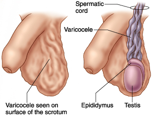
There are usually no signs or symptoms. Varicose veins sometimes cause pain, but when it is present, it can be rated as mild discomfort or acute pain. The pain may worsen if you stand or play sports for longer periods of time. Pain may be worse during the day, but will be relieved if it is in the back.
Varicocele has the opportunity to become increasingly more impressive over time. The presence of varicocele can lead to decreased sperm production, which can be remedied with the following treatments
Diagnosis and Evaluation of Varicocele
A qualified physician can make the following diagnoses varicocele Diagnosis is made by examining the patient’s situation and performing an accurate physical examination. To prove the diagnosis, varicocele ultrasound combined with a color toppler is performed. Other methods to prove the diagnosis are scintigraphy, infrared thermography, venography, and magnetic resonance imaging.
The ultrasound examination begins with the patient lying on his back with his legs spread apart. The scrotum is supported by a sling to facilitate the next exposure, while the penis is covered with a clean towel and secured to the abdomen with the help of tape.
Varicocele ultrasound With the introduction of the color jet Doppler, a Doppler is used. This is because it has the potential to literally detect the position with the highest sensitivity and specificity. A varicocele It looks like an ultrasound examination much like a worm-like tubular structure, representing a dilated vein. While the typical measuring panpini form veins is 0.5 to 1.5 mm, varicocele section, there is a chance that they vary up to 2 to 3 mm. When the patient is instructed to perform the Valsalva maneuver by breathing while the eater and mouth remain closed, the venous currents in the veins show retrograde filling. the varicocele Retrograde filling and phase changes are indicated.
Ultrasound and sonography of varicose veins
Varicocele ultrasound It actually helps to diagnose the disease by showing dilatation of the injured arteries by more than 2 mm. Valsalva maneuver or standing during the study increases venous pressure and leads to greater dilatation of these veins. Here are some pictures to demonstrate this varicocele by ultrasonography:
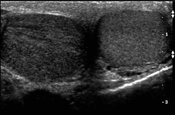
Cross-sectional scan of both test circles shows a normal left test circle compared to hypoechogenic (mild) torsion of the right test circle (Photo: Michael Bleivas, MD).
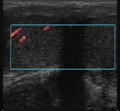
Varicocele ultrasound Kleurendoppler shows normal perfusion in the patient’s right omental tower, but the swollen left fallopian tube is not complete. (Photo Michael Bleivas, MD).
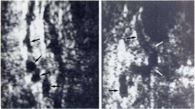
The photo on the left shows a normally messed up volume (1-1, 5 mm aperture) (arrow). The picture on the right shows a single normal vein running perpendicular (arrow) in the proximal portion of the seed chain.
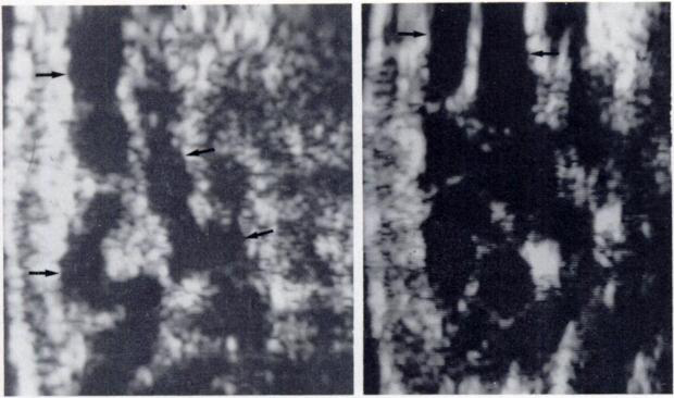
The photo on the left shows several loops of dilated veins (small.) varicocele ) of caliber within 2-3 mm. On the right side there is a large varicocele veins with a diameter of 4 to 5 mm.
Other diagnostic tools are
- CT scan helps to identify dilated clusters of serpentine veins.
- MRI may detect a varicocele Can detect dilated serpentine veins with increased blood flow.
- Venography is performed only during vascular healing. It can show dilated veins and retrograde blood flow.
Grading of varicocele Can be produced to outline the severity of the condition.
- In grade 1 varicose veins, there is no dilatation of the stray veins, but there is backflow (retrograde/reflux) into the veins of the seed chain in the gro caliber during the Valsalva’s test.
- Grade 2 varicocele shows superior veins in the upper part of the testis with reflux during the Valsalva test.
- In grade 3 varicocele, there is no non-fallus anti-inflammation in the posterior position, but in the standing position, the vein appears to be dilated in the lower part of the testicles. Reflux of blood flow is present in the veins of the inferior pole of the Valsalva test.
- Grade 4 varices also show dilation of the vein in the posterior position with reflux during the Valsalva maneuver.
- Grade 5 varicose veins show dilated veins with reflux even without Valsalva test.
What are the underlying causes?
Varicose veins are a fairly popular condition affecting about 15% of men. It usually occurs during puberty and probably leads to decreased sperm production. It is also often associated with infertility. It occurs in about 40% of men who have it.
Varicose veins are subdivided into primary and secondary type velocities.
Primary varicoceles Result of the natural ability or inability of the valves of the fallopian tubes or internal seedomer. This position usually affects the left test circle.
A secondary varicocele Result of increased pressure in the ovarian recharger. This can result from compression, obstruction, or disease related to portal hypertension.
Treatment of varicose veins
Treatment of varicocele Not always necessary unless caused by pain, decreased volume of the testicles (atrophy), or infertility.
Surgical treatment focuses on concealing the affected vein and overturning the blood flow to normal veins. In cases of infertility, healing can improve the male’s ability to produce high-quality sperm.
Risks of Repair of varicocele may include:
- Carbonized water or water accumulation around the testicles.
- Recurrence of varicocele
- Testicular atrophy (shrinkage)
- Damage to blood vessels
- Infection
Surgical repair methods include
- Open surgery under joint anesthesia or local anesthesia. In this outpatient procedure, cuts are made in the gro or gro diameter or intraperitoneally.
- Use of surgical microscopy, which improves the physician’s judgment during the procedure. Other techniques include the introduction of Doppler ultrasound as a check during the procedure. These methods are associated with the highest rate of disturbance.
- Laparoscopic surgery involves the insertion of a delicate object into a delicate bony cavity via a video camera that is supported by a video camera through a small piece of skin. This procedure is performed under general anesthesia.
- Percutaneous embolization is considered an outpatient procedure performed under local anesthesia. A long, thin tube is inserted into a vein in the gro caliber or neck and used to implement the device. The elevated vein is viewed on a monitor and the surgeon applies a solution or rinse that is interrupted to the dilated vein in the oval tower. This is where blood flow is most interrupted and the fallopian tubes are restored. the varicocele This procedure is not applied frequently.
After the procedure, it is recommended to rest for at least two days and avoid strenuous activity. Postoperative pain is usually mild and can be relieved with freely available anesthesia. Avoidance of sex is recommended for the time being.
Lifestyle and Family Relief
Not all varicoceles If surgical healing is required, remember to apply an anesthetic OTC anesthetic such as acetaminophen or ibuprofen if there is one that does not cause discomfort but does not affect fertility. You can simplify pressure by applying athletic adhealing by applying it during the work.


